Cosmetic Skin Surgery | Moles
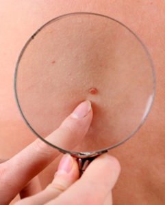
Moles or nevi are the most common pigmented skin lesions. They are composed of melanocytes and in most cases can represent an aesthetic problem.

There are many different types of moles. Depending on the genetic factors, moles can be present at birth or gradually appear later in life.
They can be different in size, color, number and pattern. Most moles that we have will not change significantly throughout years, but their appearance depends on the exposure of the skin to UV radiation.
Sometimes the size or the location of a mole can represent an inconvenience or even a difficulty in daily activities. They can be aesthetically unacceptable and can affect the self-confidence of the patient.
Persons who have moles should regularly monitor all possible changes, both by themselves and with the assistance of a dermatologist.
Early detection of mole changes and its removal is the only safe way to prevent melanoma.
When the mole is removed (regardless of the method used), a sample is sent for histopathologic examination for maximum patient safety.
How is the procedure performed?
Radiosurgery is a great method for mole removal and is widely used in plastic surgery and dermatosurgery, because it is precisely in these subspecialities of medicine essential that the procedure results with a subtle and almost invisible scar.
This method consists of removing a mole with radiofrequency current and practically evaporating the skin on the spot where radio waves are conducted. The advantages of this method are brevity, simplicity and comfort of the patient – it lasts no more than five minutes, and there are no stitches, no blood, and no pain after the intervention. After removing the mole, the scar is often more appealing than those from a surgical intervention.
The removed mole can be sent for pathohistologic analysis, and if there are any abnormal changes, this will be shown under the microscope.
Radiosurgery is also an ideal technique for all other benign changes on the skin:
warts, keratoses, fibromas, granuloma, atheromas, dermatofibromas, syringomas, csantelasmas and angiomas.
One of the greatest advantages of the radiosurgery is that it is absolutely safe for removal of changes on eyelids, with absolutely no risk for the eyes.
Radio wave interventions are very short, lasting only from 20-30 seconds, and they yield excellent aesthetic results.
Melanoma
Prognosis of melanoma depends on different factors, the most important being the growth phase, the thickness, how deeply it has grown into the skin and whether it has spread.
It also depends on several clinical indicators, so elderly people and men have poorer prognosis than younger people and women.
Of course, early stage melanoma with no metastases have better outlook. And if there is suspicion of distant metastases at the moment of diagnosis, that is very rarely the case. Fortunately, only 4% of patients have metastases at the moment when diagnosis is established.
When the mole is removed (regardless of the method used), it must be sent for histopathologic examination for maximum patient safety.
Tumors in radial phase do not have metastases, and if they have been completely removed, they do not recur.
Only 30 % of patients with metastases in regional lymph nodes have a five-year survival, while the survival rate for those with distant metastases is only 5 to 10 percent.
Since surgery is the only efficient way of treatment, the team of the Atlas hospital specialists advises regular check-up so that any potential problems may be recognized and treated on time. Recognition and early treatment of atypical moles can save life.
